Short cases in ophthalmology
Here are given the various short cases of ophthalmology that are asked from you as spotters in your MBBS practicals exams. You have to present them just as i had written. You have to identify the spot and explain it to the examiner. Also, these spotters will help you to clinically correlate the theoretical part of ophthalmology. You can download the PDF file of spotters below.Must read - Books to study ophthalmology in mbbs
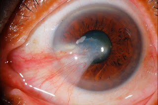
1. Pterygium
2. Stye (external hordeolum)
3. Ptosis
4. Entropion
5. Ectropion
6. Strabismus
7. Iridocyclitis
8. Retinal artery occlusion
9. Geographic corneal ulcer
10. Diabetic retinopathy
Most probably, it is the fundoscopy picture of diabetic retinopathy as it shows haemorrhages, microaneurysms and cotton wool spots.
11. Fungal corneal ulcer
This picture shows a patient with greyish white ulcer in cornea having feathery finger like extensions. A Hypopyon can also be seen. So, it is the case of fungal corneal ulcer.
12.Bacterial corneal ulcer
This picture shows a patient with descemetocele. It is formed in bacterial corneal ulcer by the bulging out of tough descemet's membrane when ulcerative process deepens.
13. Chalazion
It shows a patient of chalazion having a swelling in upper eyelid slightly away from lid margin in right eye.
14. Central retinal vein occlusion
It is the fundoscopy picture showing tomato splash appearance in retina due to massive retinal haemorrhages. Retinal viens are congested. Cotton wool spots are also present. So, it is the case of retinal vien occlusion.
It is the fundus picture of branched retinal vien occlusion as it shows venous congetion and haemorrhages that is limited to a particular area of retina.
15. Senile cataract
It is the case of morgagnian stage of senile cataract as it shows brownish nucleus of lens that had settled to bottom and also, lens had converted into bag of milky fluid.
16. Vernal keratoconjunctivitis (spring catarrh)
It shows many raised papillas in palpebral conjunctiva arranged in cobble stone fashion. It is vernal keratoconjunctivitis.
It shows whitish raised dots along the limbus called horner tranta spots.
17. Ophthalmia neonatorum
This is the case of a child having purulent conjunctival discharge coming out from both the eyes. Swelling of eyelids is also present. So, it is ophthalmia neonatorum.
18. Buphthalmos
This is a child of buphthalmos showing enlargement of eyeball. It is seen in congenital glaucoma due to the retention of aqueous humour.
19. Orbital cellulitis
It shows Proptosis along with swelling and redness of eyelids seen in orbital cellulitis.
20. Open angle glaucoma
This is the fundus photograph of a patient showing glucomatous changes in optic disc. It shows enlargement of cup and thinning of neuro retinal rim. Blood vessels are shifted nasally and giving appearance of being broken at margins ( bayonetting sign).
21 Papilloedema
It is the fundus photograph of Papilloedema showing oedema and swelling of optic disc that obliterates the physiological cup of optic disc. Engorgement of veins is also present.
22. Applanation tonometry
It is used to measure intraocular pressure. The picture in right is too small, In left is too large and In middle, it is the end point as the inner edges of 2 semicircles just touchs with each other.
Download PDF file of short cases
So, now you can easily correlate your theoritical knowledge with clinical. Thanks for reading my post and all the best for your studies.
Comment below any queries or reviews regarding this post.


~2.jpg)


8 Comments
Mbbs Admission in Ukraine follows a high standard of education and teachers here for Lowest Mbbs Fee in Russia.
ReplyDeletehttps://mbbsstudyinrussia.com/volgograd-state-medical-university/
http://mbbswisher.com/volgograd-state-medical-university/
Contact No: +91-9953971904 , 9540302883 , 7011714220
E-mail: mbbswisher@gmail.com, viptyagi83@yahoo.co.in
Great post.
ReplyDeletehttps://www.spreaker.com/user/larrywagner
Great post.
ReplyDeletehttps://www.goodreads.com/user/show/111440426-alfredo-meyer
https://www.sixsigmaedu.com/
ReplyDeleteOverseas Education Consultants, Study Abroad Counsellers in Hyderabad
Global Six Sigma Consultants, an overseas educational consultancy for students who are looking to study abroad in USA, Australia, UK, Canada, New Zealand and Europe.
overseas consultancy in hyderabad
overseas consultants in hyderabad
overseas education consultants hyderabad
8008000415
Global Six Sigma Consultants, A British based limited company exclusively offering consultation services to students intending to pursue higher studies in different Universities and colleges around the Globe. Global Six Sigma Consultants is a sister concern of Global Mobilization Societyhaving its operations for the last one decade in the field of education, capacity-building, emancipation and empowerment of youth in India and in the process of establishing its own educational academy meeting international standards.
203, Moghal Marg Building
Opp. Deepak Theatre
Himayathnagar, Hyderabad-29
Cuban MMBS medicine schools
ReplyDeleteThank you for sharing about Ophthalmology cases
ReplyDeleteBhaskar eye care clinic
Choosing a best ophthalmologist in India is challenging. Ophthalmologist is an eye doctor which diagnosis, examine your eye problems and guide you treatments or procedure accordingly your problem.
ReplyDeleteI was cured of herpes simplex virus his email ____________________________ I am so happy,
ReplyDelete-GENITAL AND ORAL HERPES
-HPV
-DIABETES
-WEAK ERECTION
-VIRGINAL PROBLEM
-WHOOPING COUGH
– HEPATITIS A,B AND C
-COLD SORE
-LOWER RESPIRATORY INFECTION
-LOW SPERM COUNT
-STAPHYLOCOCCUS AUREUS
-STROKE
-IMPOTENCE
-PILE
-HYPERTENSION
-MENOPAUSE DISEASE
-CANCER
-SHINGLES
-FIBROID
-YEAST INFECTION
-BARENESS/INFERTILITY….
Robinsonbuckler11@gmail (dot) com